The Skeletal System
What does the skeletal system do?
The skeletal system has many functions besides giving us our human shape and features.
It also:
- Allows movement: Your skeleton supports your body weight in order to help you stand and ligaments, tendons, and muscles all attach to bones, so that we can move n groove!
- Produces blood cells: Bones contain bone marrow. Red and white blood cells are produced in the bone marrow.
- Protects all of your vital organs: Your skull shields your brain, your ribs protect your heart and lungs, and your vertebrae protects your spinal cord.
- Stores minerals: Bones hold your body’s supply of minerals like calcium and vitamin D.
Did you know that the human skeleton is made up of 206 bones, including bones of the:
- Skull – (not forgetting the jawbone).
- Spine – cervical, thoracic, lumbar, sacrum and coccyx (tailbone) vertebrae.
- Chest – ribs and breastbone (sternum).
- Arms – shoulder blades (scapula), collarbone (clavicle), humerus, radius and ulna.
- Hands – wrist bones (carpals), metacarpals and phalanges.
- Pelvis – hip bones.
- Legs – thigh bones.
- Feet – tarsals, metatarsals and phalanges.
Bones
The skeleton is split into 2 main sections
Axial skeleton and Appendicular skeleton.
Have a look at the diagram below and take note of the bones in each section
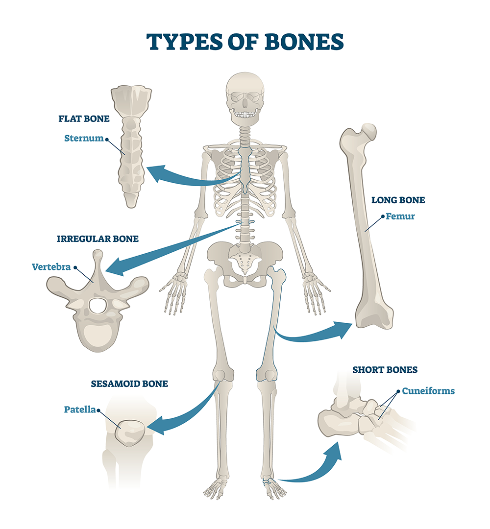
There are four different Bone types in the human body.

- Short bone – has a roughly cubed shape. Examples include the bones that make up the wrists and the ankles.
- Long bone – has a long, thin shape. Examples include the bones of the arms and legs (excluding the wrists, ankles and kneecaps).
- Irregular bone – has a shape that does not conform to any of the above. Examples include the bones of the spine (vertebrae).
- Flat bone – has a flattened, broad surface. Examples include ribs, shoulder blades, breastbone and skull bones.
- Sesamoid – are small round bones whose purpose is to reinforce and decrease stress on a tendon. Examples of where they are found are the knee, thumb, and feet.
The life story of THE BONE
Did you know that a baby grows about 10 inches in the first year of life and grows several inches each year after that, especially during puberty.
But how exactly do bones grow?
Babies are born with about 300 bones, and fully grown adults have only 206. Babies have tiny bones compared with adults and as they grow, many of these bones fuse together to create larger bones. Bones continue to grow until we reach our mid-20s, and that is when our bones will be as large as they will ever be.
Bones go through several changes as they grow. When babies are born, their bones are mostly cartilage, which is a soft and flexible substance. As babies grow, the cartilage in their bones grows. Over time and with a little help from calcium, bone replaces cartilage in a process known as ossification and simply put … ossification is a process in which bone replaces cartilage.
During ossification, layers of calcium and phosphate salts accumulate and encase the cartilage cells, and in time the encased cartilage cells die. As they die, the cartilage cells leave behind tiny pockets in the bone which are filled by blood vessels which deposit specialised cells, known as osteoblasts.
Osteoblasts are cells that deposit calcium to help form bone and produce a substance that is rich in collagen fibres that help create the structure of bone.
When the osteoblasts finish building new bone, they become flat and resemble pancakes. These flattened cells line the surface of the bone and regulate how much calcium passes into and out of the bone. Keeping the right amounts of calcium and other minerals in bone is important to maintaining strong bones.
Low calcium levels decrease bone density, which increases the risk of bone fractures.
The ossification process is typically complete by the time a person reaches their mid-20s.
So do our bones stop growing?
Not really .. the adult body replaces it’s Skeleton every 7- 10 years so after bones stop getting longer, they continue to produce new bone tissue to replace old bone tissue. Bones contain living tissue that renews itself regularly in a process known as bone turnover. The process happens in two stages. Firstly, the osteoblasts draw calcium from the bloodstream to build new bones. Next, cells known as osteoclasts dissolve the bone and return the calcium to the bloodstream.
Bone turnover continues throughout life, but the process slows down with age. During childhood and through the teen years, the body adds new bone more quickly than it removes old bone, which helps to build bone mass. After the age of about 20, the body begins to add new bone more slowly. Eventually, the body removes old bone quicker than it adds bones, which causes us to lose bone mass as we get older ; (
But there is hope … bone thickening is often in response to increased muscle activity, such as weight training. This is why weight bearing exercise .. like Body Pump etc.are fantastic classes to maintain good healthy bones particularly as you get older. Also nutrition, exposure to light and growth hormones all affect the health of the bones
Now that we have a good understanding of the life cycle of the bone, let’s take a deeper look into the long bone and see its actual structure.
Structure of a Long Bone
The long bone ‘category’ includes the legs, the arms, the metacarpals and metatarsals of the hands and feet, the phalanges of the fingers and toes, and the clavicles (collar bones).
Structure of the long bone
The outer shell of the long bone is made of compact bone. This is covered by a membrane of connective tissue called the periosteum. Beneath the compact bone (cortical bone) is a layer of spongy bone and inside this is the medullary cavity whose inner surface is lined by endosteum The medullary cavity has an inner core of bone marrow. Bone marrow is found in the inner core of the medullary cavity and has many blood vessels. There are two types of bone marrow: red and yellow. Red marrow contains blood stem cells that can become red blood cells, white blood cells, or platelets. Yellow marrow is made mostly of fat.
Interesting fact
At birth, all bone marrow is red and half of it is converted to yellow marrow by age seven.
The long bones grow primarily by elongation of the diaphysis, with an epiphysis at each end of the growing bone. When you look at the diagram you will see an epiphyseal line, this was originally an epiphyseal plate which is a thin cartilaginous line.This cartilage allows the growth of bones. Hence, it is found only in bones undergoing growth. In contrast, the epiphyseal line signifies that bone growth has stopped.
When these bones come together there is a white tissue that covers the end of the bones making it easier to move. This white tissue is called Articular Cartilage allowing the bones to glide over each other with very little friction. This Articular Cartilage can be damaged by injury or normal wear and tear.
Note: The other type of cartilage is Fibrocartilage and is a tough, very strong tissue found predominantly in the spine and at the insertions of ligaments and tendons.
So what attaches bone to bone?
Ligaments attach bone to bone and Tendons attach muscle to bone.
Ligaments and tendons are both made up of strong fibrous connective tissue, but that’s about where the similarity ends.
Ligaments appear as crisscross bands that attach bone to bone and help stabilise joints, guide joint motion, and prevent excessive motion, A ligament tear is far worse that a muscle tear as it doesn’t heal well. This is why you need to be so careful not to overload or overstretch your body. The anterior cruciate ligament (ACL) can be one of the most common tears as it attaches the thighbone to the shinbone, stabilising the knee joint.. think football, skiing … these type of sports demand movements that can really threaten the ACL
Tendons are located at each end of a muscle and attach muscle to bone. (Think of a tender muscle … tender). Tendons are found throughout the body, from the head and neck all the way down to the feet. The tendons almost blend into the muscle, so this is why it is crucial to warm up the body before a sport. For example, the achilles tendon which is the largest tendon in the body. It attaches the calf muscle to the heel bone. If you don’t do a decent warm up and have cold tight calves … OUCH!
The point where two or more bones meet to allow movement is called a joint.
Joints
How many types of joints are there in the human body?
The human body has three main types of joints. They’re categorised by the movement they allow:
Synarthroses joints (immovable). These are fixed or fibrous joints. They’re defined as two or more bones in close contact that have no movement.
Cartilaginous joints (slightly movable). These joints are defined as two or more bones held so tightly together that only limited movement can take place. Think vertebra of the spine.
Synovial joints (freely movable). These joints have synovial fluid enabling all parts of the joint to smoothly move against each other. These are the most used joints in your body so let’s go a bit deeper with these.
There are six types of synovial joints:
Synovial Joints:
- Ball and socket joint. The ball and socket joint permits movement in all directions, and features the rounded head of one bone sitting in the cup of another bone. Examples include your shoulder joint and your hip joint.
- Hinge joint. The hinge joint is like a door, opening and closing in one direction, along one plane. Examples include your elbow joint and your knee joint.
- Condyloid joint. The condyloid joint allows movement, but no rotation. Examples include your finger joints and your jaw.
- Pivot joint. The pivot joint is characterised by one bone that can swivel in a ring formed from a second bone. Examples are the joints between your ulna and radius bones that rotate your forearm, and the joint between the first and second vertebrae in your neck.
- Gliding joint. The gliding joint only permits limited movement, it’s characterized by smooth surfaces that can slip over one another. An example is the joint in your wrist.
- Saddle joint. Although the saddle joint does not allow rotation, it does enable movement back and forth and side to side. An example is the joint at the base of your thumb.
Joint movement
Still to this day …I struggle with all of the terminology in joint movement, so let’s make this as simple and easy to understand as possible.
Watch this simple, short video and hopefully it will help.
A summary of Movement Types
Movement type |
Description |
Example |
| Flexion | Decreasing the angle of a joint, or bending a limb | Bending the knee |
| Extension | Increasing the angle of a joint, or straightening a limb | Straightening the knee |
| Abduction | Taking a limb away from the mid-line of the body | Lifting the arms from the side of body |
| Adduction | Taking a limb towards the mid-line of the body | Lowering the arms towards the side of the body |
| Rotation | When a limb rotates about its own axis | Looking over your shoulder |
| Circumduction | When one end of the limb traces a circle | Doing the butterfly stroke in swimming |
| Supination | Rotation of the forearm causing the palm of the hand to face up | Turning your hands from facing down, to turning up |
| Pronation | Rotation of the forearm causing the palm of the hand to face down | Turning your hands from facing up to facing down |
| Eversion | At the ankle when the sole of the foot is turned outwards | Kicking a football with the instep |
| Inversion | At the ankle when the sole of the foot is turned inwards | When you twist your ankle, it is excessive inversion |
| Dorsi flexion | At the ankle joint when the toes are pulled upwards towards the shin | When you do a calf stretch |
| Plantar flexion | At the ankle when the toes are pointed downwards | When you stand on tip toes |
| Depression | Downward movement of the shoulder girdle | Pushing the shoulder blades down |
| Elevation | Upward movement of the shoulder girdle | Shrugging the shoulders |
| Horizontal flexion & extension | Occurs when the arm (at shoulder height) moves across the body, and back out to the side. | Bringing the arm across and then away from the body |
| Hyper extension | Excessive extension beyond straight | A crab position in gymnastics |
| Lateral flexion | Bending to the side | Tilting of the head |
| Protraction | The shoulders are drawn forwards | Rounding of the shoulders |
| Retraction | The shoulders are drawn backwards | Opening out the chest |
The Spine
Shape

Cervical .. 7 vertebrae
Allows movements of rotation, lateral flexion/extension, and flexion/extension
Thoracic 12 vertebrae
Allows movements of rotation, lateral flexion/extension and flexion/extension .. same as the cervical but in smaller ranges.
Lumbar 5 vertebra
Allows movements of rotation, lateral flexion/extension, and flexion/extension same as the thoracic but in smaller ranges.
Sacral 5 vertebrae and Coccyx 4 vertebrae ….
These are all fused and allow no movement.
So, the lower you go the less mobile you are!
The spine plays a HUGE role in stability and movement of the body and protects the spinal cord which is the guy that sends all those important messages to and from the brain.
If you look at the spine from the side you’ll notice the way the spine curves in (concave) and out(convex). Think of the man IN his cave ..concave. It’s this S shape that enables the spine to function properly as the core to our balance and stability.
Bones of the spine
The Spine is comprised of 33 Irregular bones called the vertebra and has 5 different regions. Each region have a different number of bones which are shaped differently to allow movement and impact.
The Neutral Spine
When you start to teach, you’ll hear again and again the term “Neutral Spine”
Neutral spine is the natural position of the spine when all 3 curves of the spine — cervical, thoracic and lumbar — are present and in good alignment.
This is the strongest position for the spine when we are standing or sitting.
It’s ideal that we have our spine in a neutral position as our back and neck will not be placed under stress and strain … but in this day and age .. this doesn’t happen naturally hence the popularity of classes like Pilates that really work on educating clients about the importance of a neutral spine and strong core
Common poor posture habits can create abnormalities in the spine
Lordosis
This is when the abdominal muscles are long and weak and the back extensor muscles are tight and short .. the result .. a hollow back appearance
Kyphosis
This is when the muscles at the front of the chest and upper back are shortened and the mid back muscles are lengthened .. the result .. a hunched back appearance.
Flat back
This is when you lower back flattens out, losing the curve in your spine and tipping the pelvis backward.
Sway back
Looks really like Lordosis but Lordosis is caused by an over curvature of the spine, whereas sway back is when the shoulder is behind the pelvis.
Scoliosis
This is when there is an excessive bend to one side of the spine and the body will try and compensate to control and stabilise the spine.
Watch this short video that summerises Body Types
Poor posture can also be the result of exercise or sport imbalances. think fencing, golfing, .. one side works much harder than the other.
Age – As we get older, our shape changes and our bones can deteriorate. This can also be due to medical conditions such as osteoporosis, spina bifida and cerebral palsy. The latter two conditions can have a massive impact of the formation of the spine.
Note; These are topics that you will definitely want to cover at a late date.
So why do you need to know about the Skeletal System?
Now that you are training to become a purestretch Instructor, you need to know the positive impact exercise can have on the Skeletal System.
Here are just a few:
- Increases bone density and strength, so less chance of fractures.
- Reduces the risk of osteoporosis.
- Increases your range of movement, so a better performance.
- Improves your posture.
- Improves the health of cartilage, so less tear and wear on those bones.
The next Chapter is all about the Neuromuscluar Systems but before we go there let’s just have a quick revise and see what you remember?


